초음파 검사의 혜택과 응용
초음파 검사, 또는 초음파 영상은 현대 의학 진단과 모니터링에 중요한 역할을 합니다. 고주파 음파를 사용하여 신체 내부의 이미지를 생성하는 초음파 검사는 많은 혜택을 제공하며 다양한 건강 상태를 진단하고 관리하는 데 필수적입니다. 여기에서는 초음파의 주요 장점과 이 기술로 가장 많은 혜택을 받는 특정 질병과 건강 상태를 살펴봅니다.
초음파 검사의 주요 혜택
비침습적이고 통증 없음: 초음파는 절개가 필요하지 않은 비침습적 절차로, 환자에게 통증이 없고 안전합니다.
방사선 없음: X선 및 CT 스캔과 달리 초음파는 이온화 방사선을 사용하지 않습니다. 이는 특히 임산부와 태아에게 안전한 옵션이 됩니다.
실시간 이미지: 초음파는 실시간 이미지를 제공하여 상태가 발생하는 즉시 진단하고 모니터링하는 데 매우 중요합니다. 이는 특히 산과에서 태아의 실시간 모니터링이 필요한 경우에 중요합니다.
다양한 응용 분야: 초음파는 임신 모니터링, 간, 신장 및 심장과 같은 장기 상태 진단, 생검과 같은 절차 안내, 혈관의 혈류 평가 등 다양한 의학적 목적으로 사용됩니다.
접근성 및 편리성: 초음파 기기는 클리닉, 병원, 원격 지역을 포함한 다양한 환경에서 사용할 수 있습니다. 절차는 보통 15-45분 정도로 짧고 특별한 준비가 거의 필요하지 않습니다.
비용 효율성: MRI 및 CT 스캔과 같은 다른 영상 기술에 비해 초음파는 일반적으로 비용이 적게 들어 환자와 의료 제공자 모두에게 비용 효율적인 옵션입니다.
이상 발견: 초음파는 연조직, 장기 및 혈관의 종양, 낭종 및 기타 병변을 발견하는 데 도움을 줍니다. 조기 발견은 효과적인 치료와 관리에 중요할 수 있습니다.
치료 진행 모니터링: 초음파는 다양한 상태에 대한 치료 진행 상황을 모니터링하여 치료가 효과적인지 확인하고 필요한 경우 조정할 수 있습니다.
초음파 검사로 가장 많은 혜택을 받는 건강 상태
초음파 검사는 다양한 질병과 건강 상태를 진단하고 모니터링하는 데 매우 유익합니다. 여기에는 다음이 포함됩니다:
임신 및 산과
태아 발달: 태아 성장 모니터링, 선천적 이상 감지, 태아 건강 상태 평가.
태반 및 양수: 태반의 위치 평가 및 양수 양 측정.
다태 임신: 쌍둥이나 다태 임신 관리 및 모니터링.
복부 상태
담낭 질환: 담석, 염증 또는 기타 담낭 문제 감지.
간 질환: 간 종양, 낭종, 지방간, 간경변 및 기타 간 이상 감지.
신장 질환: 신장 결석, 낭종, 종양 감지 및 신장 기능 평가.
췌장 상태: 췌장염, 종양 또는 기타 췌장 이상 진단.
대동맥류: 복부 대동맥류 감지 및 모니터링.
골반 상태
부인과 문제: 난소 낭종, 자궁근종, 자궁내막증 및 기타 자궁 상태 진단.
전립선 질환: 전립선 크기 평가 및 종양이나 비대와 같은 이상 감지.
방광 문제: 방광 종양, 결석 또는 기타 이상 감지.
심혈관 상태
심장 질환: 심장 기능 평가, 판막 장애 감지 및 심장 구조 평가를 위한 심장 초음파 검사.
혈관 상태: 혈류 평가, 혈전 감지(심부정맥 혈전증), 말초 동맥 질환 평가.
유방 상태
유방 종괴: 고형 종괴와 낭종 구별, 생검 안내 및 유방 임플란트 평가.
갑상선 질환
갑상선 결절: 결절, 낭종 및 기타 갑상선 이상 감지 및 평가.
갑상선종: 갑상선 크기 및 구조 평가.
근골격계 상태
관절 및 연조직 손상: 인대 파열, 근육 손상, 건염 및 기타 연조직 이상 진단.
류마티스 질환: 관절 염증 평가 및 관절염과 같은 상태에 대한 주사 안내.
소아 상태
선천적 이상: 신장, 심장 및 뇌와 같은 장기의 선천적 이상 감지.
유문 협착증: 유아의 유문 협착증 진단.
감염성 질환
농양: 농양 감지 및 배농.
림프절 비대: 림프절 비대 평가를 통해 감염과 악성 종양 구별.
결론
초음파 검사는 현대 의학 진단과 환자 관리에서 다재다능하고 안전하며 효과적인 도구입니다. 방사선 관련 위험 없이 실시간으로 상세한 이미지를 제공함으로써 초음파는 다양한 건강 상태의 진단 및 관리에 매우 중요합니다. 접근성, 비용 효율성 및 광범위한 응용 가능성으로 인해 초음파 검사는 조기 발견, 정확한 진단 및 수많은 건강 문제의 효과적인 모니터링을 보장하는 현대 의학 실습의 필수 구성 요소입니다.
The Benefits and Applications of Ultrasound Checkups
Ultrasound checkups, also known as sonograms, play a vital role in modern medical diagnostics and monitoring. Utilizing high-frequency sound waves to create images of the inside of the body, ultrasound exams offer numerous benefits and are integral in diagnosing and managing a variety of health conditions. Here, we explore the key advantages of ultrasound and the specific diseases and health conditions that benefit most from this technology.
Key Benefits of Ultrasound Checkups
Non-invasive and Painless: Ultrasound is a non-invasive procedure that does not require incisions, making it painless and safe for patients.
No Radiation: Unlike X-rays and CT scans, ultrasounds do not use ionizing radiation. This makes them a safer option, particularly for pregnant women and developing fetuses.
Real-time Imaging: Ultrasound provides real-time images, which can be crucial for diagnosing and monitoring conditions as they happen. This is especially important in obstetrics, where real-time monitoring of the fetus is necessary.
Wide Range of Applications: Ultrasound is used for a variety of medical purposes, including monitoring pregnancy, diagnosing conditions in organs such as the liver, kidneys, and heart, guiding procedures like needle biopsies, and evaluating blood flow in vessels.
Accessibility and Convenience: Ultrasound machines are widely available and can be used in various settings, including clinics, hospitals, and even remote locations. The procedure is quick, typically taking only 15-45 minutes, and requires little to no special preparation.
Cost-effective: Compared to other imaging techniques like MRI and CT scans, ultrasounds are generally less expensive, making them a cost-effective option for both patients and healthcare providers.
Detecting Abnormalities: Ultrasounds can help detect abnormalities such as tumors, cysts, and other pathologies in soft tissues, organs, and blood vessels. This early detection can be critical for effective treatment and management.
Monitoring Treatment Progress: Ultrasound can be used to monitor the progress of treatment for various conditions, ensuring that therapies are effective and making adjustments if necessary.
Health Conditions Benefiting Most from Ultrasound Exams
Ultrasound exams are highly beneficial for diagnosing and monitoring a wide range of diseases and health conditions, including:
Pregnancy and Obstetrics
Fetal Development: Monitoring fetal growth, detecting congenital anomalies, and assessing fetal well-being.
Placenta and Amniotic Fluid: Evaluating the position of the placenta and measuring amniotic fluid levels.
Multiple Pregnancies: Managing and monitoring twin or multiple pregnancies.
Abdominal Conditions
Gallbladder Disease: Detecting gallstones, inflammation, or other gallbladder issues.
Liver Disease: Identifying liver tumors, cysts, fatty liver, cirrhosis, and other liver abnormalities.
Kidney Disorders: Detecting kidney stones, cysts, tumors, and evaluating kidney function.
Pancreatic Conditions: Diagnosing pancreatitis, tumors, or other pancreatic abnormalities.
Aortic Aneurysms: Identifying and monitoring abdominal aortic aneurysms.
Pelvic Conditions
Gynecological Issues: Diagnosing ovarian cysts, fibroids, endometriosis, and other uterine conditions.
Prostate Disorders: Evaluating prostate size and detecting abnormalities like tumors or enlargement.
Bladder Problems: Detecting bladder tumors, stones, or other abnormalities.
Cardiovascular Conditions
Heart Disease: Evaluating heart function, detecting valve disorders, and assessing heart structures using echocardiography.
Vascular Conditions: Assessing blood flow, detecting blood clots (deep vein thrombosis), and evaluating peripheral artery disease.
Breast Conditions
Breast Lumps: Differentiating between solid masses and cysts, guiding biopsies, and evaluating breast implants.
Thyroid Disorders
Thyroid Nodules: Identifying and evaluating nodules, cysts, and other thyroid abnormalities.
Goiter: Assessing the size and structure of the thyroid gland.
Musculoskeletal Conditions
Joint and Soft Tissue Injuries: Diagnosing ligament tears, muscle injuries, tendonitis, and other soft tissue abnormalities.
Rheumatologic Conditions: Evaluating joint inflammation and guiding injections for conditions like arthritis.
Pediatric Conditions
Congenital Abnormalities: Detecting congenital abnormalities in organs such as the kidneys, heart, and brain.
Pyloric Stenosis: Diagnosing pyloric stenosis in infants.
Infectious Diseases
Abscesses: Identifying and draining abscesses.
Lymphadenopathy: Evaluating enlarged lymph nodes to differentiate between infection and malignancy.
Ultrasound checkups are a versatile, safe, and effective tool in medical diagnostics and patient care. By providing real-time, detailed images without the risks associated with radiation, ultrasounds are invaluable in diagnosing and managing a wide array of health conditions. Their accessibility, cost-effectiveness, and broad applicability make them an essential component of modern medical practice, ensuring early detection, accurate diagnosis, and effective monitoring of numerous health issues.
위암에 대해
문경미 박사님께서 매주 건강 관련 블로그를 게시할 계획입니다. 궁금한 사항이 있거나 글을 읽고 이해가 되지 않는 부분이 있다면 언제든 댓글을 남겨주세요. 여러분과 소통하며 건강한 삶을 이루고자 합니다. 함께 동참해주시길 바랍니다.
위에 발생하는 악성 종양에는 여러 종류가 있습니다. 대표적으로는 위 선암, 림프종, 위 점막하 종양, 평활 근육종 등이 있죠. 그 중에서도 위 선암이 약 98%를 차지하는데, 이에 따라 우리가 '위암'이라고 말할 때는 보통 위 선암을 의미합니다.
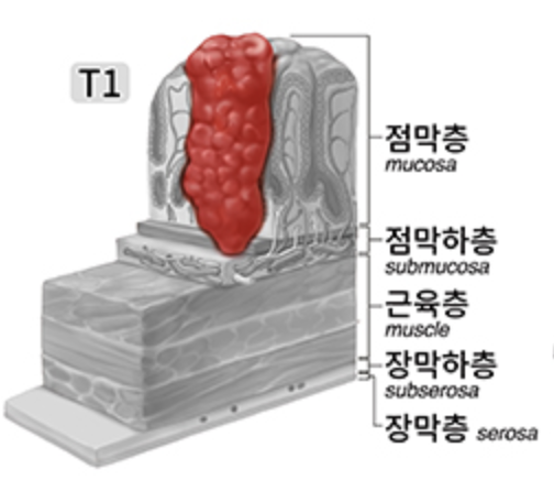
위암은 위의 점막에서 발생합니다. 시간이 지나면서 종양은 점막하층, 근육층, 장막하층, 그리고 장막층까지 확산합니다. 때로는 종양이 점막이나 점막하층을 따라 위 내부로 넓게 퍼지기도 하며, 점막층에서 장막층으로까지 깊이 침투하기도 합니다. 또한 종양은 위 주변의 임파선을 따라서, 혹은 혈류를 통해 간, 폐, 뼈 등 다른 부위로 퍼질 수 있다는 점에서 매우 위험합니다. 그러나 조기 위암 단계에서 수술하게 되면 완치가 가능합니다.
위암은 여러 요인에 의해 발생할 수 있습니다.
만성 위축성 위염: 위암의 높은 위험 요소 중 하나로, 위암으로 진행할 위험성이 높은 전구병변입니다. 위암으로 진행되기까지 걸리는 시간은 약 16~24년 정도입니다.
장 이형성: 위암의 전 단계 병변으로 알려진 현상으로, 위점막 세포의 소장 선세포가 나타나는 것을 말합니다.
위소장 문합술: 위와 소장을 연결하는 수술 후 위 산도가 낮아지면 세균이 증식하고 박테리아가 집중됩니다. 이로 인해 위암 발병 위험도가 20년 후에 3~5배 높아집니다.
식이 요인: 질산염 화합물과 고염 식품, 그리고 불에 구운 음식, 술, 담배 등이 위암 발병 위험을 높일 수 있습니다. 반면에 채소, 과일, 비타민 등은 항암 효과가 있습니다.
헬리코박터 파일로리균(Helicobacter pylori) 감염: 이균에 감염되면 위암 발병 위험도가 증가할 수 있습니다.
유전 요인: 유전적으로 선종성 대장폴립을 가진 사람이나 가족력에 위암이나 대장암이 있는 경우 위암 발병 위험이 높아집니다.
환경적 요인: 석면, 철가루 먼지, 공해, 전리방사선, 흡연, 방부제, 농약, 산업폐기물 등이 위암을 유발하는데 영향을 줄 수 있습니다. 불행히도, 위암은 대부분 초기에는 별다른 증상을 보이지 않을 수 있습니다.
위암의 증상으로는 상복부 불쾌감, 상복부 통증, 소화불량, 팽만감, 식욕 부진 등이 나타날 수 있습니다. 그러나 이러한 증상들은 종종 위염이나 위궤양과 유사합니다. 이로 인해 많은 사람들이 소화제나 제산제를 장기간 복용하며 대증 요법을 시도하며 수술의 시기를 놓치는 경우가 있습니다. 조기에 위암을 찾아내기란 쉽지 않기 때문에 정기적인 검사가 꼭 필요합니다. 특히 60세 이상의 경우, 증세가 미약할지라도 보름 이상 위장 질환이 지속될 경우 반드시 검사를 해야합니다.
위암이 조기에 발견되지 않으면 증상이 점차 악화되어 복부에 단단한 덩어리가 만져질 수 있습니다. 또한 구토, 토혈, 하혈, 체중 감소, 빈혈, 복부의 부어오름 등의 증상이 나타날 수 있습니다. 이런 경우에는 종종 암이 진행된 정도로 치료 결과가 좋지 않을 수 있습니다.
위암은 증상과 진찰만으로는 진단하기 어렵습니다. 그러나 복부 초음파나 위 내시경을 통해 진단할 수 있으며, CT 및 조직 검사를 통해 최종 진단이 이루어집니다. 위 내시경 검사는 조금 불편할 수 있지만, 위장 내부를 직접 확인하고 의심되는 부위에 대해 조직 검사를 할 수 있어 위암과 헬리코박터 파일로리균(Helicobacter pylori) 감염 여부를 동시에 확인할 수 있는 장점이 있습니다. 최근에는 수면 내시경 검사로 인해 검사에 따른 불편함이 줄어들었습니다.
점막층에 국한된 조기 위암의 경우 수술로 95% 이상의 완치 효과를 기대할 수 있습니다. 그러나 진행성 위암은 5년 생존율이 30% 내외에 불과하여, 예후가 매우 나쁩니다. 그런데 위장 검사를 받으면 위암으로 인한 사망을 50%까지 감소시킬 수 있다는 연구 결과가 보고되었습니다. 위암의 근본적인 예방이 현실적으로 불가능한 현재로서는, 조기에 위암을 찾아내 치료하는 것이 최선입니다.
다음에는 위암의 치료 방법에 대해 알아보겠습니다.


